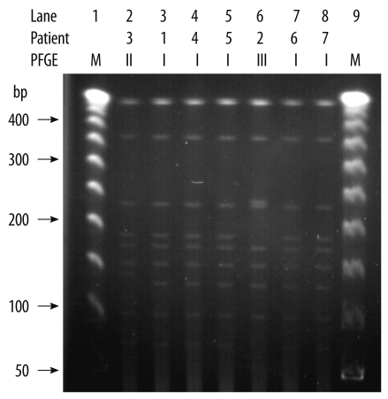
Fig. 1.
Pulsed-field gel electrophoresis (PFGE) patterns of SmaI-digested genomic DNA obtained from the ribotype 027 isolates. Lanes 1 and 9 were loaded with a 50-kb ladder (molecular marker). The PFGE patterns for isolates 2 and 3 differed in 5 bands. The remaining 5 isolates had identical patterns, and their pattern differed from that of isolates 2 and 3 in 3 and 2 bands, respectively.

