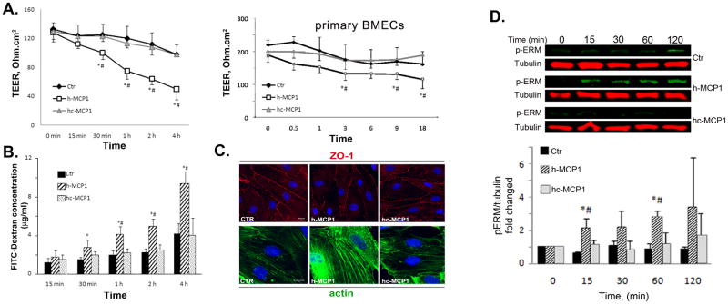
Figure 4. Mouse MCP1 C-terminus inhibits human MCP1-induced BBB compromise.
A. Left panel: 100 nM h- or hc-MCP1 was added to the lower chamber of the bEND.3-Astrocyte co-culture system. Right panel: Primary BMEC monolayer was treated with 100 nM h- or hc-MCP1 (Primary BMECs). TEER values were determined over time in the presence of A2AP. Values are mean ± SD (n=3–4). TEER values were determined over time in the presence of A2AP. Values are mean ± SD (n=4). B. Cells were treated as in A. The leakage of FITC-Dextran across the in vitro BBB was determined by a fluorescence plate reader in the presence of A2AP. The concentration of FITC-Dextran in the lower chamber was calculated based on a standard curve. Values are mean ± SD (n=4). Analysis was performed using student’s t-test. Comparison with Ctr, *p<0.05 and comparison with hc-MCP1, #p<0.05. C. Mouse MCP1 C-terminus inhibits human MCP1-induced redistribution of tight junction proteins. Primary BMEC were treated with 100 nM h- or hc-MCP1 for 2 hours in the presence of A2AP. The cells were then fixed and immunostained for ZO-1 (upper panels) and F-actin (lower panels). D. Mouse MCP1 C-terminus is inhibitory to human MCP1-induced phosphorylation of ERM proteins. bEND.3 cells were treated with saline or 100nM h- or hc-MCP1 proteins in the presence of A2AP over time. The phosphorylated ERM proteins were determined using anti-phosphor-ERM antibody. Quantitative data of western blots are shown as mean ± SD (n=3). Comparison with control, *p<0.05 and comparison with hc-MCP1, #p<0.05.

