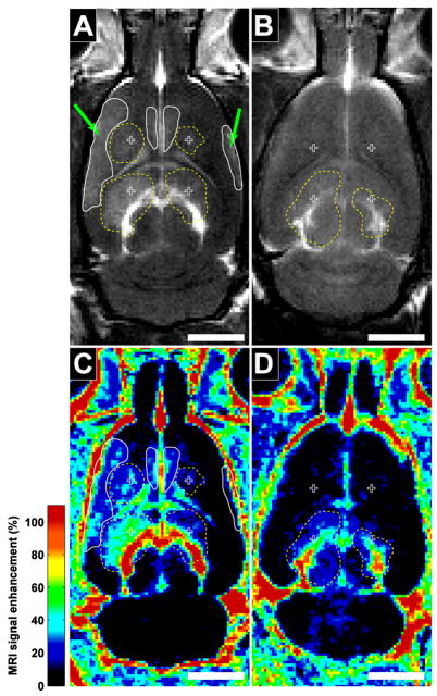
Figure 3.
Examples of contrast-enhanced T1-weighted FSE images in the rat brain after sonication with an unfocused transducer in animals under ketamine/xylazine (A) and isoflurane/oxygen (B) anesthesia. Four locations were targeted in each animal (crosses). Left locations were sonicated at 4.1 W and right locations at 2.8 W (corresponding to peak pressures in water of 250 and 230 kPa). MRI contrast-enhancement at the sonication targets (dotted outlines) is clearly evident for animals anesthetized with ketamine/xylazine at all four locations. With isoflurane/oxygen, only minor enhancement at the two inferior locations was observed. With ketamine/xylazine, additional enhancement away from the targets (solid outlines) was also evident at peripheral brain regions (arrows) and near the midline, presumably from reflections or standing waves within the cranium. (bar: 5 mm)

