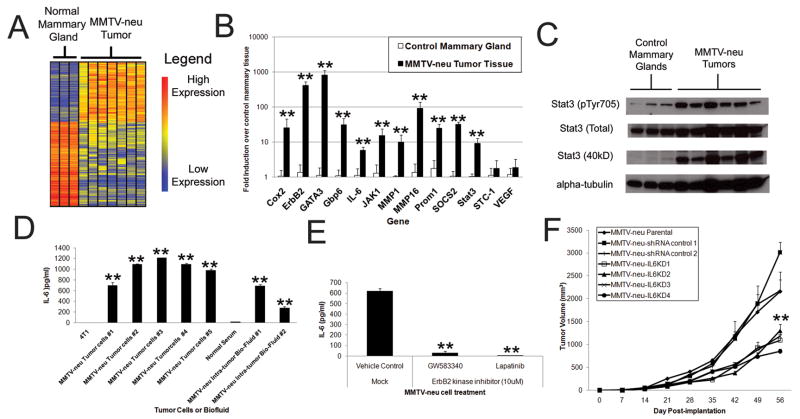
Figure 6. ErbB2-mediated induction of IL-6 plays a critical role in an endogenous ErbB2-mediated model of breast cancer.
A) Heatmap depiction of significantly dysregulated genes (1-way ANOVA with p<.05, >3 fold difference) in MMTV-neu tumors. B) Quantitative rt-PCR from MMTV-neu tumors and control mammary glands (N=5, bars indicate SD). C) Western blot analysis of lysates of MMTV-neu tumors and control mammary glands. D) Tumor cells from MMTV-neu tumors were isolated and cultured assess IL-6 secretion in comparison to a non-ErbB2 transformed mammary carcinoma cell line (at 24 hours post-plating). Fluid samples from tumors as well as control mice serum were also tested for IL-6 concentration by ELISA. In all samples N=3, bars represent SD. E) Tumor cells from MMTV-neu mice were passaged for 3 months, mock or ErbB2 kinase inhibitor treated, and IL-6 secretion assessed at 24 hours post-treatment (N=3, bars indicate SD). F) MMTV-neu tumor cells were modified by IL6KD or control lentiviral infection and implanted (1×10⁶ cells) via subcutaneous injection into SCID mice and tumor volume measured over time (N=5, bars represent SE). Where indicated * and ** represent p<.05 and p<.01 respectively in comparison to controls.

