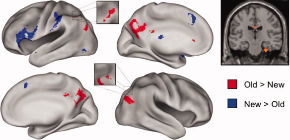
Figure 1.

Left: Regions demonstrating old > new and new > old effects common to the recall and recognition tasks are displayed in red and blue, respectively, on the left and right lateral and medial hemispheres of an inflated fiducial brain (see Methods). Right: The common old > new effect in the right medial temporal lobe is displayed on a section at y = −20 through the canonical single‐subject T1‐weighted image. Effects are thresholded for display purposes at P < 0.0025.
