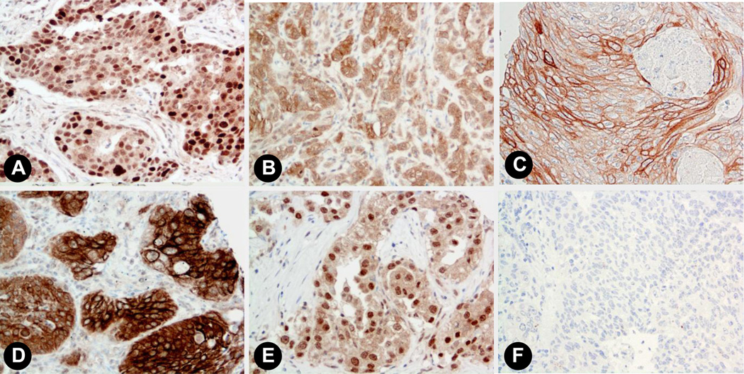
Fig. 3. Progesterone receptor expression in human lung tissues.
Representative images showing staining patterns for progesterone receptor in normal lung and NSCLC (20x magnification). A) Breast cancer positive control. B) Adenocarcinoma showing relatively stronger extranuclear as compared to nuclear staining. C) Squamous cell carcinoma showing strong membrane staining; D) Squamous carcinoma with some of the tumor cells showing both intense membrane and cytoplasmic staining. E) Adenocarcinoma showing considerably stronger nuclear staining relative to extranuclear staining. F) Non-immune antibody-incubated negative controls showed no staining.

