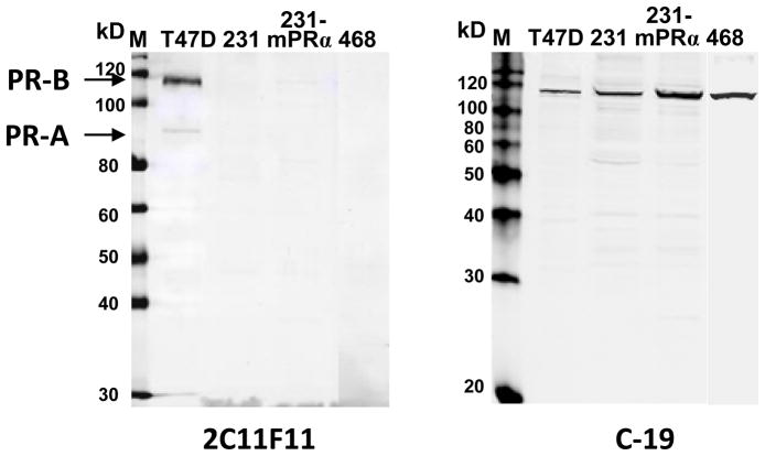
Fig. 2.
Western blot analyses of PR-positive and PR-negative breast cancer cell lines using two C-terminal PR antibodies. M: prestained (left) and Western (right) protein size marker; T47D: T47D Yb cell lysate; 231: MDA-MB-231 cell lysate; 231-mPRα: mPRα transfected MDA-MB-231 cell lysate; 468: MDA-MB-468 cell lysate. Sample proteins: 40μg/lane. Left panel, detected with 2C11F11 anti PR C-terminus antibody from Santa Cruz. Right panel, detected with C-19 antibody from same source.

