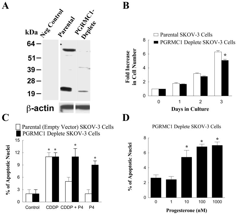
Figure 2.
The development of a dsRed-SKOV-3 cell line that was depleted in PGRMC1. Panel A is a western blot showing PGRMC1 levels in parental and PGRMC1-deplete dsRed SKOV-3 cells. β-actin western blot is shown to demonstrate equal protein loading. The − sign indicates a lane of a western blot in which the primary antibody was omitted and replaced with IgG (i.e. a negative control). The rate of cell proliferation of parental and PGRMC1-deplete SKOV-3 cells is shown in panel B. The effect of cisplatin (CDDP; 30 nM) and progesterone (P4; 1 μM) on the rate of apoptosis of parental empty vector and PGRMC1-deplete SKOV-3 cells is shown in panel C. The effect of increasing concentrations of P4 on the percentage of PGRMC1-deplete SKOV-3 cells undergoing apoptosis is shown in panel D. Data taken from Peluso et al [11].

