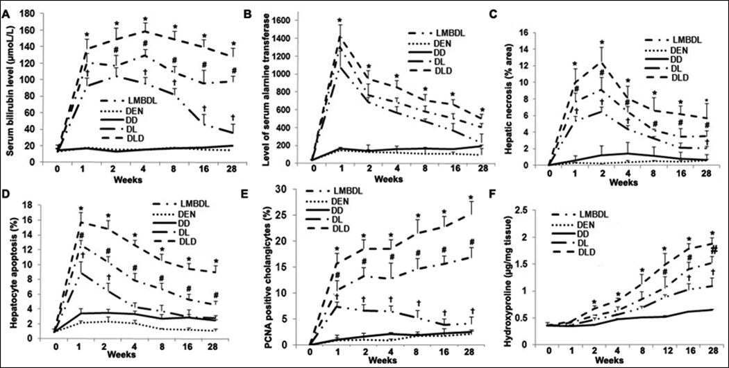
Fig. 2. Assessment of liver damage and histopathological features in the ligated lobes of the treated mice.

Total serum was extracted from the treated groups to assess A) total bilirubin and B) ALT levels. C) Hepatic necrosis was determined by H&E staining. D) Hepatocyte apoptosis was identified by TUNEL staining. E) Cholangiocyte proliferation was evaluated by PCNA staining. F) Hydroxyproline content was done to look at fibrosis. Five liver tissue samples for each group were used for the above assays. *p<0.05 DLD vs all other groups, #p<0.05 DL vs LMBDL, †p<0.01 LMBDL vs DEN and DD groups.
