Fig. 6.

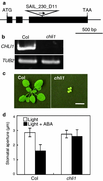
Characterization of the T-DNA insertion mutant for Mg-chelatase I subunit 1 (chli1). a Schematic representation of the structure of the CHLI gene and the location of the T-DNA insertion site. Black boxes and lines represent exons and introns, respectively. The T-DNA was inserted into the third exon of the CHLI1 genomic DNA. b CHLI1 expression verification by RT-PCR in the chli1 mutant (upper panel). TUB2 was used as a control (lower panel). c Four-week-old plants, Col and chli1, grown under a 16-h photoperiod. Bar 10 mm. d Effect of ABA on stomatal closing in Col and chli1 plants. Stomatal apertures were measured after incubation for 2.5 h in the basal buffer with (black) or without (white) 20 μM ABA. Other conditions were the same as in Fig. 1d. Data represent the mean with SD. Experiments repeated three times on different occasions gave similar results
