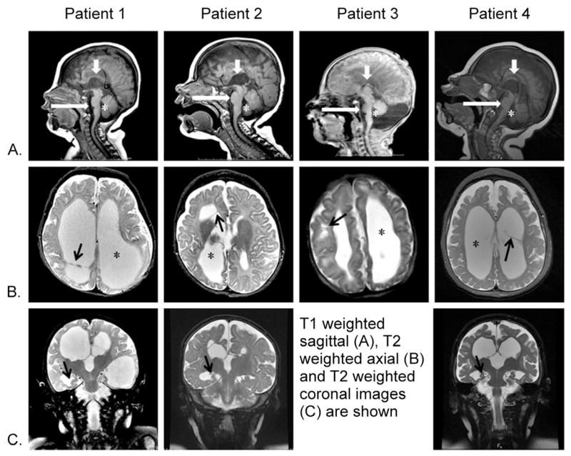
Figure 1. MRI characteristics in 4 female patients with PDHA1 deficiency.
Columns show patients 1, 2, 3, and 4 imaged at 4 months, 5 months, 1 day, and 11 months, respectively. Rows: A, sagittal midline T1-weighted images; B, axial T2-weighted images at the level of lateral ventricles; C, coronal T2-weighted images through the hippocampi.
Note the incomplete corpus callosum in all patients (Row A, short white arrows). All patients had asymmetrical ventriculomegaly (Row B, stars) with a normal 4th ventricle (Row A, stars). A small pons (Row A, long white arrows) can be seen. Ventricular septations were seen in 3/4 patients (row B, patients 1,2 and 4, black arrows) or parenchymal cysts in 1/4 patients (row B, patient 3, black arrow). Patients 1,2 and 4 displayed hyporotated hippocampi (row C, arrows).

