FIGURE 1.
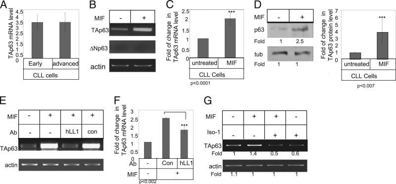
p63 is a target gene of CD74 in CLL cells. A, Early- and advanced-stage B cells derived from CLL patients were purified. Quantitative real-time PCR was performed using primers for TAp63 and RP-2, as described in Materials and Methods. B–D, CLL cells were incubated in the presence or absence of MIF. B and C, After 18 h, RNA was purified. B, TAp63, ΔNp63, and actin mRNA levels were analyzed by RT-PCR. n = 23 patients. C, Quantitative RT-PCR: results are expressed as a fold of change in TAp63 expression by stimulated cells compared with nonstimulated cells, which was defined as 1. n = 5 patients. D, Cells were lysed after 24 h exposure to MIF, and p63 and tubulin expression were analyzed by Western blot analysis. Graph summarizes the results of four different experiments. E and F, CLL cells were incubated in the presence or absence of MIF, hLL1, or a control Ab for 18 h. E, RNA was purified, and levels of TAp63 and actin mRNA were analyzed. The results presented are representative of six CLL patients. F, Quantitative RT-PCR: results are expressed as a fold of change in TAp63 expression in stimulated cells compared with nonstimulated cells, which was defined as 1. Results shown are a summary of three separate experiments. G, CLL cells were incubated in the presence or absence of MIF or ISO-1 for 18 h. RNA was purified, and levels of TAp63 and actin mRNA were analyzed. The results presented are representative of five CLL patients.

