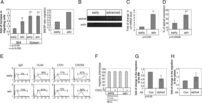
FIGURE 2.
Migration of advanced CLL cells to the BM is enhanced due to elevation of VLA-4 integrin. A, Early CLL cells stained with CFSE and advanced CLL cells stained with CMTMR were injected i.v. into C57BL/6 mice. BM and spleen populations were analyzed by FACS after 3 h. The graph presents the averages of four early and three advanced CLL patients. B and C, Early- and advanced-stage B cells derived from CLL patients were purified, and RNA was purified. B, VLA-4 and actin (10 early and 15 advanced CLL patients) mRNA were analyzed. C, Quantitative RT-PCR was performed using primers for α4 integrin (11 early and 9 advanced) and RP-2 (as described in Materials and Methods). D, Cells were stained with anti-α4 and analyzed by FACS. Graph shows α4 integrin (17 advanced and 21 early) expression on CLL cells. E, Cells were stained with anti–LFA-1, anti-α4, CXCR4, and CD19 and analyzed by FACS. Histograms show the expression of various receptors on CD19-positive cells. The results presented are representative of two early and two advanced CLL patients. F, CLL cells accumulated at 0.75 dyn/cm2 for 60 s on HUVECs alone, or HUVECs overlaid with CXCL12 were subjected to physiological shear stress (5 dyn/cm2) for 15 min. Results show relative numbers of each migratory phenotype. The graph is representative of two early (VLA-4 and LFA-1 low) and two advanced (VLA-4 high and LFA-1 low) patients. G and H, CLL cells were incubated in the presence of anti–VLA-4 or a control Ab for 30 min. Cells were stained with CFSE and were injected i.v. into C57BL/6 mice for 3 h. Bone marrow (G) and spleen (H) populations were analyzed by FACS. n = 3. TEM, transendothelial migration.

