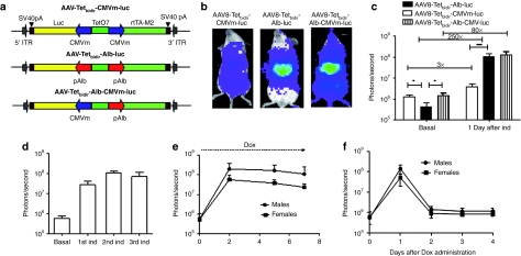
Figure 1.
In vivo characterization of AAV-pTetbidir-CMV-luc and AAV-pTetbidir-Alb-luc. (a) Schematic diagram of tetracycline-inducible adeno-associated viral (AAV) vectors used in this study. Alb, albumin promoter; CMVm, minimal early cytomegalovirus promoter; ITR, inverted terminal repeat; luc, luciferase gene; polyA, SV40 fragment containing the early and late polyadenylation signals; rtTAM2, mutated reverse tetracycline transactivator; TetO7, sequence-containing seven tetracycline operator sites. (b) BALB/c mice injected with 5 × 10¹² vg/kg of AAV8-Tetbidir-CMVm-luc, AAV-Tetbidir-Alb-luc or AAV8-Tetbidir-Alb-CMVm-luc were analyzed with the charge-coupled device (CCD) camera before and 24 hours after administration of doxycycline (Dox). Shown are optical CCD images for luciferase expression of representative animal from each group, 1 day postinduction. (c) Luciferase expression was quantified and represented as photons/second. The data are shown as mean ± SD. The differences on luciferase expression was statistically evaluated by Student's t-test (*P < 0.05, ***P < 0.001). (d) Mice injected with AAV-Tetbidir-Alb-luciferase were reinduced 15, 30, and 60 days after vector injection and luciferase expression was measured. (e) AAV8-Tetbidir-Alb-luc, male and female BALB/c mice were injected intravenous (i.v.) with a dose of 5 × 10¹² vg/kg. Two weeks after injection, luciferase expression was induced by a single intraperitoneal (i.p.) injection of Dox and expression induction was maintained by continuous administration of Dox in drinking water over the course of a week. Each dot represents the average of the decimal logarithm of bioluminescence for each group of animals at a time point. (f) The extinction time of luciferase expression was determined after removal of Dox.

