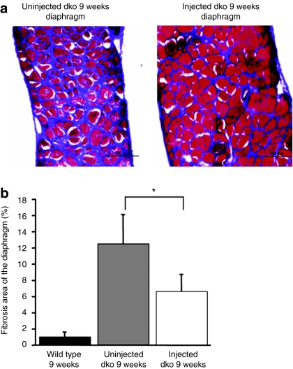
Figure 4.
Reduction of secondary degeneration by exogenous dystrophin. (a) Decrease in fibrotic area in the injected dko diaphragm. The Masson trichrome staining reveals interstitial fibrosis (aniline blue stained). Masson trichrome-stained transverse sections of 9-week-old dko mice (left; uninjected, right; injected) were shown. Bar = 100 µm. (b) Fibrotic area of injected dko mice showed a significant decrease compared with uninjected dko mice (n = 3–4 mice/group wild type: 1.03 ± 0.26%, uninjected dko: 12.5 ± 2.3%, injected dko: 6.7 ± 0.81%, uninjected dko versus injected dko: *P < 0.05).

