
Figure 4. Inflammation in KI mice is induced by IL-1β.
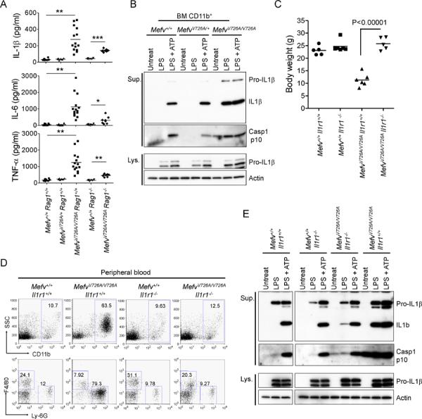
(A) Luminex analysis of serum cytokines of 8-week-old WT, MefvV726A/+, MefvV726A/V726A Rag1−/−, and MefvV726A/V726A Rag1−/− mice. *, P < 0.05; **, P < 0.0001; ***, P < 0.000001. (B) CD11b+ cells from BM of WT, MefvV726A/+, and MefvV726A/V726A mice were stimulated with LPS or no stimulus for 3 hr followed by treatment with or without ATP. Cell culture supernatants (Sup.) and cell lysates (Lys.) were analyzed by immunoblotting. (C–E) Analyses of MefvV726A/V726A mice on the Il1r1−/− background. (C) Body weights of 8-week-old males. (D) Flow cytometry analyses of peripheral blood cells from 8-week-old mice. Data are representative of three independent experiments. (E) Inflammasome activation in MefvV726A/V726A mice withIl1r1−/− background. Culture supernatants and lysates from BM CD11b+ cells were collected and subjected to immunoblot as described in (B). Additional information is provided in Figure S3.