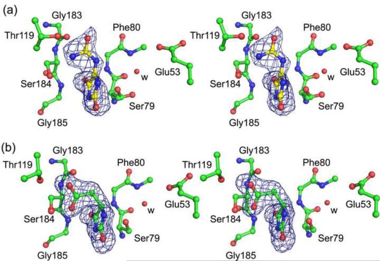
Fig. 3.
Ligands bound to KpHpxA. (a) Stereodiagram of allantoin bound to the C79S/C184S KpHpxA double mutant. (b) Stereodiagram of 5-acetylhydantoin bound to the C79S/C184S KpHpxA double mutant. In both cases carbon atoms are green, oxygen atoms are red and nitrogen atoms are blue. The red sphere labeled with 'w' is a water molecule. The sidechain of Phe80 has been omitted for clarity. The electron density in both cases is from difference maps generated before the addition of the ligand molecule into the model and are contoured at 3σ.

