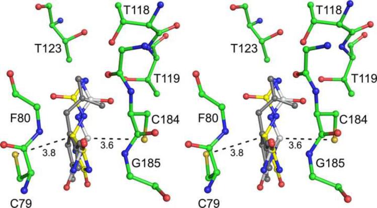
Fig. 6.
Allantoin isomers in KpHpxA active site. Stereodiagram of the (R)- (white carbon atoms) and the (S)- (yellow carbon atoms) isomers of allantoin docked in the active site of KpHpxA. The observed distances between the two active site cysteines and C5 of the respective modeled allantoin isomers are shown in Ångstroms. The observed enol form of allantoin is shown in grey.

