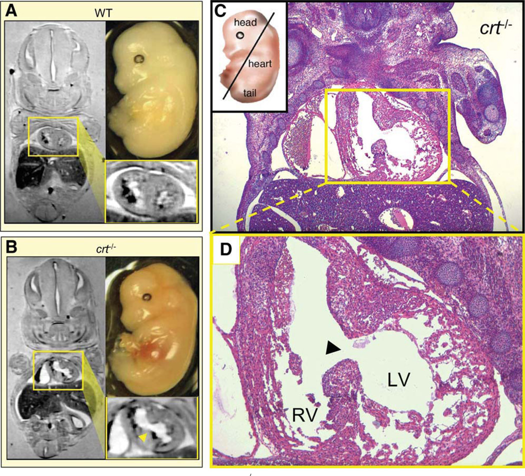
Figure 7.
Magnetic resonance reveals ventricular septal defect in crt−/− embryo. Comparative in situ magnetic resonance imaging of coronal planes from 14.5 dpc-old WT and crt−/− embryos demonstrates normal (A) versus defective (B) cardiac structure, respectively, as a predicted manifestation of cardiac phenotype. Magnified cardiac digital cross-section (yellow boxes) shows prominent ventricular septal defect with interventricular communication in crt−/− (arrowhead) in contrast to distinct left and right ventricular lumens in WT hearts characteristic for this stage of cardiogenesis. (C): Pathoanatomical verification of crt−/− dysmorphic cardiac structure (right). Coronal hematoxylin-eosin stained thin section of embryo visualizes a direct communication (ventricular septal defect) between LV and RV through a patent ventricular septum in the crt-null mutant. (D): Magnification of cardiac section highlights incomplete septal development, a common manifestation of congenital heart disease. Histology confirms known defect of a thin left ventricular myocardium characterized by deep intertrabecular recesses with the predominance of large fenestrations. Right ventricle demonstrates normal muscularization without pathological thinning or gross abnormalities. Abbreviations: LV, left ventricle; RV, right ventricle; WT, wild type.

