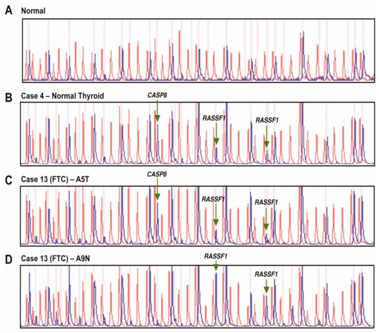
Figure 2.
(A) Normal control DNA sample with 41 individual peaks (red) in the absence of HhaI and 15 separate peaks (blue) in the presence of HhaI. (B) Normal thyroid sample with methylation of CASP8 and RASSF1. (C) Follicular thyroid cancer (Case 13 - tumor block) with methylation of CASP8 and RASSF1. (D) Follicular thyroid cancer (Case 13 -normal block) with methylation of RASSF1. (FTC – follicular thyroid cancer, A5T – block A5 tumor, A9N – block A9 normal).

