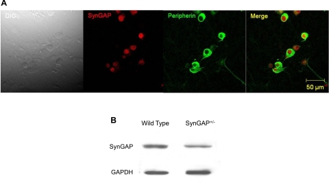
Fig. 1.
Expression of synaptic GTPase-activating protein (SynGAP) in peripheral sensory neurons. A: immunofluorescent staining of dissociated dorsal root ganglia (DRG) sensory neurons [from wild-type (WT) mice] showing that SynGAP (red) is present in neurons, also marked with the neuronal marker, peripherin (green). The horizontal bar represents 50 μm, and the magnification is 40×. DIC, differential interference contrast. B: immunoblots of DRG extracts showing SynGAP expression in WT and heterozygous mutation of the SynGAP gene (SynGAP+/−) mice.

