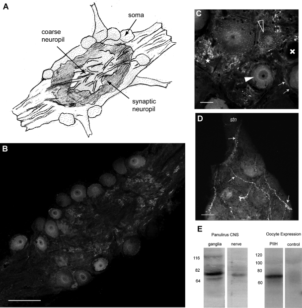
Fig. 1.
Panulirus interruptus hyperpolarization-activated inward current (Ih) protein (PIIH)-like immunolabeling is located in the somata and in the neuropil regions throughout the stomatogastric ganglion (STG). A: general anatomy of the STG. The STG contains about 30 motor and interneurons. The somata are situated on the outer surface of the ganglion and send large neurites toward the coarse neuropil in the middle, from where smaller processes branch into the layer of the fine (or synaptic) neuropil, located between the somata and the coarse neuropil. B: PIIH-like immunolabeling is found in STG somata and in the fine neuropil, whereas the larger processes of the coarse neuropil in the middle of the ganglion are much less brightly stained. Single 1-μm-thick optical section of the confocal stack. Scale bar, 150 μm. C: variable PIIH-like immunolabeling in and around the STG somata. Most neurons show strong perinuclear (filled arrowhead) or punctate staining in the soma (open arrowheads), whereas some are apparently lacking PIIH-like immunolabeling (cross). Dense PIIH-like immunolabeling is present in tissue surrounding the somata in the form of strong punctate staining (star) and halo-like layers (small arrows) around cell bodies, which may be glia or connective tissue. Single 1-μm-thick optical section of the confocal stack. Scale bar, 30 μm. D: PIIH-like immunolabeling reveals large fibrous structures (arrows) of unknown origin, which enter the STG from the stomatogatric nerve (stn) and dorsal ventricular nerve and branch within the ganglion. Maximum intensity projection of 7 consecutive confocal sections. Scale bar, 30 μm. E: specificity of the PIIH antibody was determined with Western blots of Panulirus central nervous system (CNS) tissue and from Xenopus oocyte, expressing PIIH RNA. The antibody labeled a strong (∼76 kDa) band in protein extracted from ganglia and from stomatogastric nervous system nerves (left). A larger band in the range of 116 kDa was found in Panulirus brain tissue and may be phosphorylated, glycosylated, or oligomeric forms, whereas the faint smaller band (∼64 kDa) may be the result of protein breakdown or smaller splice variants. Comparison of protein extracted from Xenopus oocytes without (control) and with injection of PIIH RNA shows a strong PIIH-positive band of similar weight in the PIIH-expressing oocytes. The Panulirus tissue and the oocyte extracts were run separately with different molecular mass ladders.

