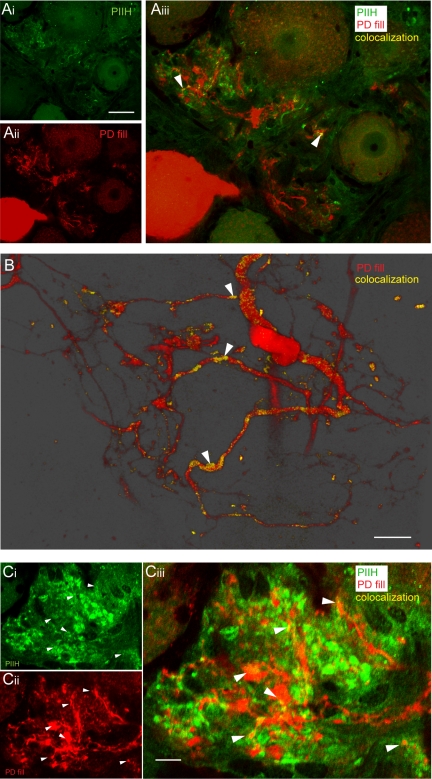
Fig. 4.
PIIH-like immunoreactivity in the pyloric dilator (PD) fine neuropil. A: overlay of PIIH-like immunolabeling in the fine neuropil of a sectioned ganglion (Ai) with a Lucifer yellow-labeled PD neuron (Aii) revealed relatively sparse PIIH-like immunolabeling of the PD neuropil (arrowheads in Aiii). Scale bar, 50 μm. B: a minority of branches of the PD neuron showed more extensive PIIH-like immunolabeling, as shown in a 3-D rendered transparency projection from 25 1-μm-thick optical slices from a different preparation. Scale bar, 30 μm. C: clusters of PIIH-like immunolabeling in the fine neuropil (Ci) occurred in close proximity to the dye-labeled PD neuropil (Cii), with occasional overlap visible (filled arrowheads in Ciii). Single optical section (0.45 μm) of a sectioned ganglion. Scale bar, 10 μm.

