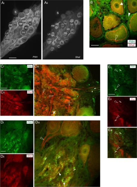
Fig. 6.
PIIH-like and Shal immunoreactivity in the STG. A: overview of PIIH-like (Ai) and Shal immunoreactivity (Aii) in the same STG whole mount shows a distinctly different labeling pattern. Whereas PIIH-like immunoreactivity was found throughout the STG in neurons and the surrounding tissue, Shal immunoreactivity was concentrated in neurons with high concentrations in the primary neurites. Scale bar, 150 μm. AB, anterior burster neuron; AM, anterior median neuron; DG, dorsal gastric neuron; GM, gastric mill neuron; LG, lateral gastric neuron; MG, median gastric neuron; PY, pyloric neuron; S, STG. B: PIIH-like and Shal immunolabeling overlap in the soma of STG neurons. Both show a perinuclear distribution of labeling (arrow), but only Shal was highly concentrated in the soma membranes (open arrowhead), and only PIIH-like immunolabeling was found in the tissue surrounding the soma (filled arrowhead). Scale bar, 30 μm. C: PIIH-like (Ci) and Shal immunolabeling (Cii) in the coarse neuropil. Transparency projection of 7 1.3-μm optical slices reveals cloudy and patchy PIIH-like immunoreactivity throughout the coarse neuropil (Ci), whereas anti-Shal distinctly labeled large processes of the primary neurites (Cii). Overlay shows that large processes in the coarse neuropil generally lack PIIH-like immunoreactivity while showing strong Shal immunolabeling (open arrowheads in Cii). Some PIIH-like immunolabel was also found in or in close vicinity of axons, shown for anterior lateral nerve (arrows, right) on leaving the STG. Scale bar, 70 μm. D: single optical section shows area of the fine neuropil, which shows intense PIIH-like (Di) and Shal labeling (Dii). At a superficial level, both signals occurred within the same regions of the fine neuropil (Diii). Note the complete absence of PIIH-like immunoreactivity in the first stretch of the primary neurites, where the most intense Shal immunolabeling was found (open arrowhead). In the neuropil, PIIH-like staining is punctate (flat arrowheads) and only occasionally appears to overlap with Shal immunolabeling (filled arrowheads). Scale bar, 30 μm. E: at high magnification, PIIH-like labeling (Ei) and Shal immunolabeling (Eii) are frequently found in close apposition throughout the fine neuropil but rarely are overlapping and colocalizing. At this level of magnification, PIIH-like immunolabeling still appears concentrated in a patchy or punctate pattern (flattened arrowheads in Eiii), which is found in the close vicinity of Shal-positive structures (open arrowheads in Eiii), but the overlay shows that both signals rarely overlap at the pixel range (Eiii). Scale bar, 20 μm.

