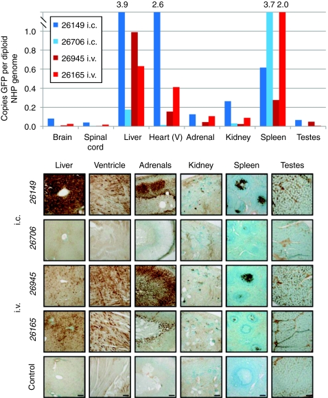
Figure 9.
Nonhuman primate (NHP) biodistribution and expression in peripheral organs. At the time of euthanasia, various peripheral organs were rapidly dissected and frozen in liquid nitrogen for quantitative PCR (qPCR) analysis of vector genome biodistribution (top panel) or postfixed in 4% paraformaldehyde and processed for immunohistochemical detection of green fluorescent protein (GFP) by 3,3′-diaminobenzidine tetrachloride staining (bottom panel). For the qPCR biodistribution studies, the “brain” is the average value of three samples and the “spinal cord” is the average of cervical and lumbar samples. Bottom: animals with no pre-existing antibodies (i.c., 26149; i.v., 26945) showed high levels of transduction in the liver, heart, and adrenals whereas the kidney, spleen, and testes showed lower levels of GFP expression. The animal with pre-existing neutralizing antibodies (NAbs) (i.c., 26706) had no observable GFP expression except in the spleen. The fourth NHP showed rising levels of NAbs throughout the 4 weeks postadministration and had high expression of GFP in the liver, moderate in the heart and adrenals, and low in the kidney, spleen and testes. Scale bar for each organ is shown in the picture of the control sample. Bar = 100 µm.

