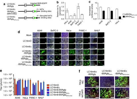
Figure 3.
Construction and characterization of recombinant microRNA (miRNA)-regulated vaccinia virus (MRVV). (a) Schematic representation of the recombinant vaccinia virus genome showing the modified B5R protein fused with enhanced green fluorescent protein at its C-terminus (MRVV/BG). Four copies of let-7a miRNA complementary or disrupted target sequences, flanked by NheI/AgeI restriction sites, were incorporated into the 3′-untranslated region of the B5R gene. (b) Relative expression of mature let-7a miRNA in the indicated cell lines by real-time PCR analysis. The data are the let-7a level normalized with the U6 small nuclear RNA level relative to that in HeLa cells and are represented by the mean ± SD (n = 3). (c) The cell lines expressing different levels of let-7a were transfected with pMirGlolet7a-mut or pMirGlolet7a plasmid containing two expression units encoding firefly luciferase (FLuc) used as the primary reporter to monitor mRNA regulation and Renilla luciferase (RLuc) acting as a transfection control. Dual luciferase assay was performed 24 hours post-transfection. The FLuc activity is normalized to the RLuc activity. The data are presented as mean + SD (n = 3). (d) The cell lines expressing different levels of let-7a were infected with the MRVV/BG at an multiplicity of infection (MOI) of 0.1 and photographed using phase-contrast or fluorescence microscopy of the same field 3 days later. Bar = 200 µm. (e) One-step growth of the MRVV/BG was determined by titration of the viruses that were collected from the infected cells shown in (d). The data are presented as mean + SD (n = 3). (f) HeLa-let7aKD cells (let-7a miRNA knockdown) or HeLa-NC cells (negative control) were infected with the MRVV/BG at an MOI of 0.1 and photographed 3 days later. The combined phase-contrast and fluorescence images are shown. Bar = 200 µm.

