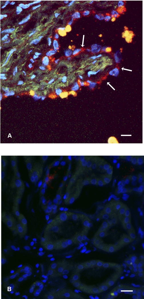
Fig. 2.
Cadherin 8 expression in autosomal dominant polycystic kidney disease (ADPKD) and normal kidney. A: tissue section from an ADPKD kidney, labeled with anti-cadherin 8 (red, arrows) and Hoescht 33342 (blue) to label nuclei. Matrix staining in green is the result of an autofluorescent signal. Note that the cyst-lining epithelial cells express cadherin 8 predominantly in a cytoplasmic distribution (arrows). Cadherin 8 may have redistributed owing to prolonged cross-clamp time required during the nephrectomy. Bar, 20 μm. B: tissue section from a normal human kidney labeled with anti-cadherin 8 (red) and Hoescht 33342 (blue) to label nuclei. Green channel is the result of an auto fluorescent signal. No cellular staining is noted. Bar, 20 μm.

