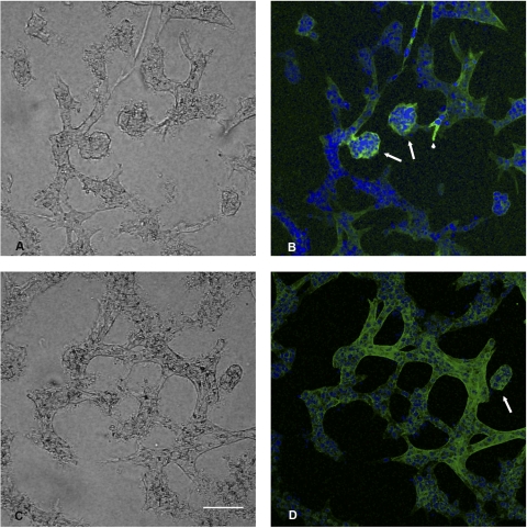
Fig. 4.
Immune fluorescence images of cysts staining for cadherin 8 or N-cadherin. 3D matrix culture was fixed with paraformaldehyde 48 h after adenovirus microinjection with both cadherin 8 and tet trans-activating adenovirus. A: phase-contrast image of B. B: extended focus image of image stack in which the sample was stained with anti human cadherin 8. Note the cystic outgrowths (arrows) and a small early stalk (arrowhead) expressing cadherin 8 (FITC label-green). Hoescht 33342 (blue) staining of nuclei is also shown. C: phase-contrast image of D. D: N-cadherin staining (FITC label-green) is observed in tubule structures and a cyst structure (arrow). Hoescht 33342 (blue) staining of nuclei is also shown. Bar, 135 μm.

