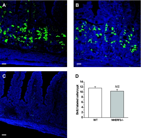
Fig. 2.
Cell proliferation in ileum from NHERF2−/− mice is not different from WT. A–C: 5-bromo-1-(2-deoxy-β-d-ribofuranosyl) uracil (BrdU) incorporation (green) was quantitated after 2 h of labeling in crypt cells of WT (A), NHERF2−/− ileum (B), and ileal tissue from NHERF2−/− injected with PBS as control (C). D: morphometry of BrdU-incorporated cells 2 h after injection in WT and NHERF2−/− ileal tissue revealed no difference in number of cells stained with anti-BrdU antibodies. Scale bars in A–C, 20 μm.

