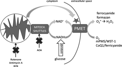
Fig. 7.
Model for the role of PMET in β-cells. PMET is measured by the transplasma membrane reduction of couplers [1-methoxy-5-methyl-phenazinium methyl sulfate (mPMS) or CoQ1], followed by the reduction of membrane-impermeable indicator dyes (WST-1 or ferricyanide). The activity of the PMET system is proportional to the intracellular concentration of NADH. Glucose undergoes glycolysis, increasing the cytosolic concentration of NADH, resulting in PMET stimulation. Application of mitochondrial inhibitors of complex I, III, and IV prevents mitochondrial oxidation of NADH, stimulating a rise in cytosolic NADH, which also results in enhanced PMET activity. Similarly, treatment with the malate/aspartate shuttle inhibitor AOA blocks mitochondrial-mediated reoxidation of NADH, leading to enhanced PMET activity.

