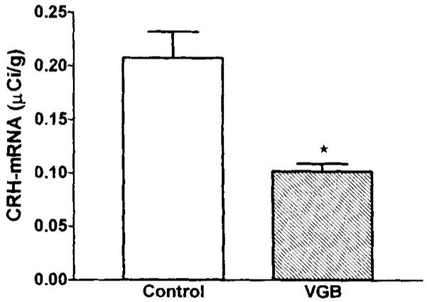
FIG. 2.

CRH–mRNA levels in the hypothalamic paraventricular nucleus (PVN) are decreased after VGB treatment. Quantitative analysis of autoradiograms of coronal sections (six per brain) at the level of the hypothalamus subjected to in situ hybridization for CRH–mRNA. Signal over the paraventricular nucleus (PVN) was quantified by using densitometric analysis. Values are expressed as mean ± SEM of 18–24 values per group. Significance of difference from vehicle-treated controls was determined by using the unpaired t test with Welch’s correction: *p < 0.001.
