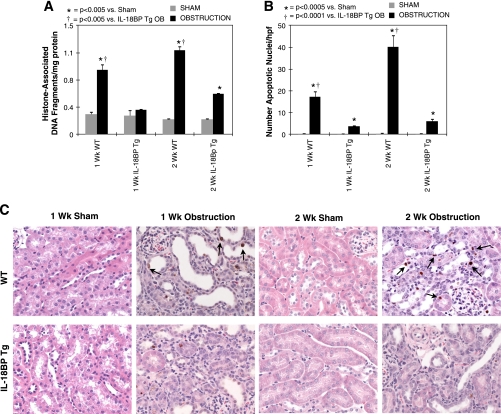
Fig. 1.
Renal cell apoptosis following unilateral ureteral obstruction (UUO). A: renal cortical histone-associated DNA fragment accumulation (ELISA) in wild-type (WT) and IL-18-binding protein transgenic (IL-18BP Tg) mice exposed to sham operation or 1 or 2 wk of UUO. B: graph depicting the number of apoptotic nuclei (TdT-mediated dUTP nick end labeling) per high-powered field (×400) in each treatment group. C: photographs (×400) depicting apoptotic nuclei (ApopTag) in renal cortical tissue sections counterstained with hematoxylin and eosin from WT and IL-18BP Tg mice exposed to sham operation or 1 or 2 wk of UUO. Apoptotic nuclei are stained brown and identified with black arrows.

