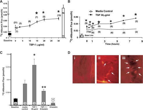
Fig. 1.
Dose-, time-, and protein tyrosine kinase (PTK)-dependent effects of thrombospondin-1 (TSP1) on transendothelial 14C-albumin flux. Postconfluent human lung microvascular endothelial cells (HMVEC-L) monolayers were exposed to increasing concentrations of TSP1 or medium alone for increasing exposure times. A: ●, mean (±SE) 14C-BSA flux in pmol/h immediately after 6-h exposures to increasing concentrations of TSP1. B: circles represent mean (±SE) 14C-BSA flux in pmol/h immediately after increasing exposure times to TSP1 (30 μg/ml, i.e., 214 nM) (●) or medium alone (○). C: vertical bars represent mean (±SE) transendothelial 14C-BSA flux in pmol/h immediately after 6-h exposures to medium alone, TSP1 (30 μg/ml, i.e., 214 nM), TSP1 + erbstatin (5 μM), or erbstatin alone. In A, B, and C, mean (±SE) pretreatment baseline transendothelial 14C-BSA flux is shown by the closed bars and flux across naked filters by the open bar (A); n indicates number of monolayers studied and is indicated next to each symbol (A + B) or within each bar (C). *Significantly increased compared with the simultaneous medium control at P < 0.05. **Significantly decreased compared with TSP1 alone at P < 0.05. D: postconfluent HMVEC-L monolayers were exposed for 6 h to TSP1 (30 μg/ml, i.e., 214 nM) (ii and iii) or medium alone (i), after which they were probed with the F-actin probe, rhodamine-phalloidin, and processed for fluorescence microscopy. Arrows indicate intercellular gaps in TSP1-exposed monolayers. Magnification = ×800.

