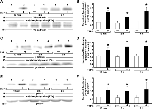
Fig. 3.
Tyrosine phosphorylation of zonula adherens (ZA) proteins over time. HMVEC-Ls were incubated with TSP1 (30 μg/ml, i.e., 214 nM) or medium alone for 10 min, 2 h, or 6 h in the presence of vanadate (200 μM) and PAO (1.0 μM) only during the last 10 min of incubation. At each time point, ECs were lysed and the lysates immunoprecipitated with antibodies raised against VE-cadherin (A), γ-catenin (C), or p120ctn (E), and each immunoprecipitate was processed for phosphotyrosine immunoblotting. To control for protein immunoprecipitation, loading, and transfer, blots were stripped and reprobed with each immunoprecipitating antibody. Each blot is representative of 3 independent experiments. B, D, and F: densitometry for each ZA phosphotyrosine signal was normalized to the densitometry for the respective total ZA protein signal in the same lane in the same gel. B: normalized densitometry for VE-cadherin (n = 3). D: normalized densitometry for γ-catenin (n = 3). F: normalized densitometry for p120ctn (n = 3). *Significantly increased compared with the simultaneous medium control at P < 0.05.

