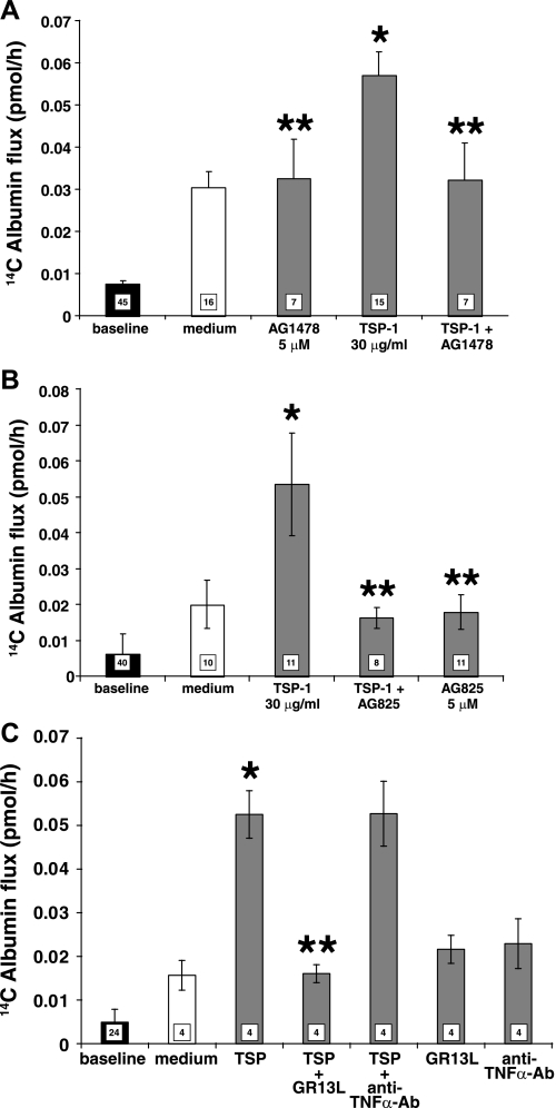
Fig. 6.
TSP1 opens the endothelial paracellular pathway through EGFR/ErbB2 activation. HMVEC-Ls cultured to postconfluence in barrier assay chambers were exposed for 6 h to TSP1 (30 μg/ml, i.e., 214 nM) or medium alone in the presence or absence of the EGFR-selective tyrphostin, AG1478 (5 μM) (A), the ErbB2-selective tyrphostin, AG825 (5 μM) (B), or the GRI3L antibody that blocks the ligand-binding portion of the EGFR ectodomain or a species- and isotype-matched antibody control (C). Vertical bars represent mean (± SE) transendothelial 14C-BSA flux in pmol/h immediately after the 6-h study period. The mean (± SE) pretreatment baselines are shown by the closed bars; n, the number of monolayers studied, is indicated in each bar. *Significantly increased compared with the simultaneous media control at P < 0.05; **significantly decreased compared with TSP1 alone at P < 0.05.

