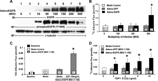
Fig. 9.
EGFR overexpression in HMVEC-Ls increases EC responsiveness for TSP1 EGF-like repeats. Adenovirus encoding for human EGFR (Ad-EGFR) or green fluorescent protein (GFP) (Ad-GFP) were packaged and amplified in human embryonic kidney 293 cells. A: HMVEC-Ls cultured to confluence in 60-mm dishes were incubated with media alone or infected with packaged Ad-EGFR at 105-108 plaque forming units per dish, i.e., at approximate multiplicities of infection (MOIs) of 1, 10, 50, 100, 150, 200, and 500, respectively. At 24 h, the cells were lysed and the lysates processed for immunoblotting for EGFR. The blots were stripped and reprobed for phosphoEGFR (Y1068) and again for β-tubulin to indicate protein loading and transfer. B: HMVEC-Ls were cultured to postconfluence in barrier assay chambers, after which they were infected with either Ad-EGFR or Ad-GFP at increasing MOIs (0–200), after which transendothelial 14C-BSA flux was measured. C and D: postconfluent HMVEC-Ls were infected with Ad-EGFR or Ad-GFP at an MOI of 150. After the baseline barrier integrity for each infected monolayer was established, the cells were exposed for 6 h to EGF (100 ng/ml) (C), increasing concentrations of recombinant TSP1 EGF-like repeats (D), or medium alone (C and D), after which barrier function was assayed. *Significantly increased compared with either the simultaneous Ad-GFP (B and D) or medium (C) controls at P < 0.05.

