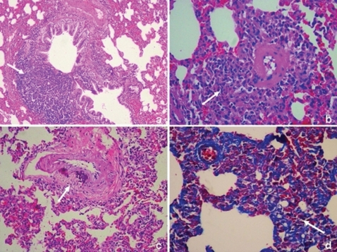
Figure 3.
Histopathological findings in the cigarette smoke-exposed group (Group II). (a) peribronchial inflammation (arrow) (H&E, ×20). (b) perivascular inflammation and vascular wall thickness (arrow) (H&E, ×20). (c) vascular wall thickness and calcification (arrow) (H&E, ×20). (d) parenchymal infiltration and fibrosis (arrow) (Masson's trichrome, ×20).

