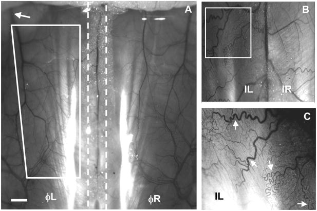
Figure 2.
Intravital microscopy images showing perfused (functional) vasculature (6.25x) in the mouse spinotrapezius muscle. A) The vasculature of the mouse spinotrapezius muscles on day 0 located to the left and right of the spine (indicated by the dashed lines). The approximate ischemic zone can be seen bounded by the four white lines. Sutures to induce the ischemic injury were placed around the feeder arteriole (approximate location upstream of white arrow) as it exited laterally from the spinotrapezius muscle. Scale bar is 1 mm. B) Intravital image digitally zoomed and enhanced. Mice that received ischemic ligations exhibited pronounced tortuosity and increased vascular branching five days post-surgery. The large vein in the center is the spinal vein, with either spinotrapezius to the left (ischemic) or right (contralateral control). Note the differing vascular architectures and pronounced vessel tortuosity on the ischemic side (white box). C) Digitally zoomed (white box) and image-enhanced view of small arterioles and venules, almost down to the capillary-level (additional digital zoom not undertaken during quantification of microvascular remodeling). A high degree of vessel tortuosity is evident (white arrows).

