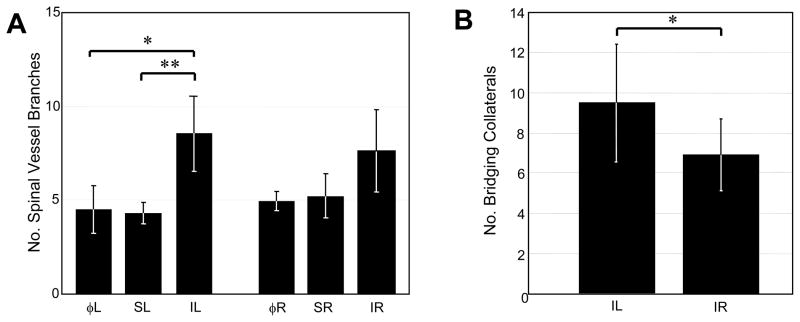
Figure 4.
Intravital microscopy. A) Quantification of spinal artery and vein branches five days post-arterial ligation (n=6), sham ligation (n=3), or at day 0 prior to any intervention (n=6). Bars correspond to muscle groups analyzed (see Methods). B) Quantification of bridging collaterals in tissues receiving ischemic ligation (IL) and their paired contralateral controls (IR) five days post-arterial ligation (n=6). Vessels were considered bridging collaterals if they crossed within 1 mm of spinal artery-vein pair, independent of origin. Values are mean ± SD *p ≤ 0.05.

