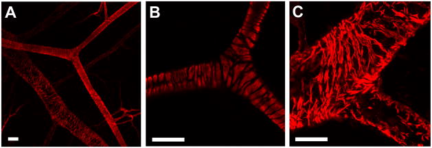
Figure 7.
Whole-mount immunolabeling of spinotrapezius muscles for αSMA using confocal microscopy. (A) Tight wrapping of VSMCs is visible on the arterioles, while a loose cobblestone morphology is evident on the venules (20x). (B) 60x image of bifurcating arteriole. (C) 60x image of venule. Note the high-level of cellular detail accessible by confocal microscopy in this model. Scale bars are 50 μm.

