Figure 1.
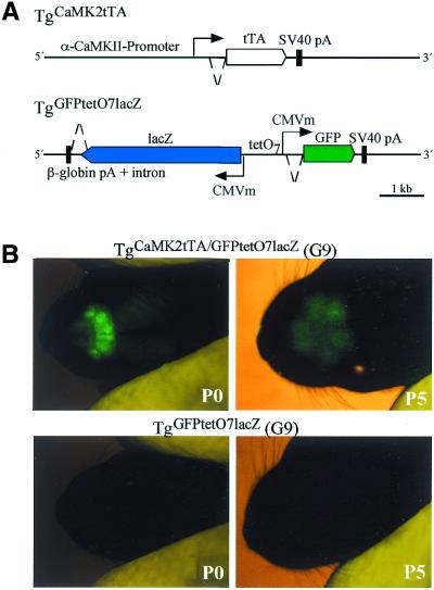
α-CaMKII-promoter controlled GFP expression in the mouse. (A) Schematic drawing of transgenes for regulated GFP and β-galactosidase expression . The upper line shows the tTA activator transgene from line TgCaMK2tTA and the lower line describes the tTA-responsive minigenes used to produce lines G3, 7, 8 and 9 (TgGFPtetO7lacZ). CMVm shows the position of transcriptional start sites after tTA has bound to the TetO7. Dashed lines, intronic sequences; arrows, transcriptional starts, black boxes, transcriptional stops; open arrows, open reading frames. (B) Stereomicroscopic identification of GFP expressing TgCaMK2tTA/GFPtetO7lacZ mice compared to non-activated TgGFPtetO7lacZ living pups of line G9 at P0 and P5. The overview shows GFP fluorescence in regions of the olfactory epithelium, the olfactory bulb and the cortex. At P5, exposure time was increased 4-fold to monitor GFP fluorescence.

