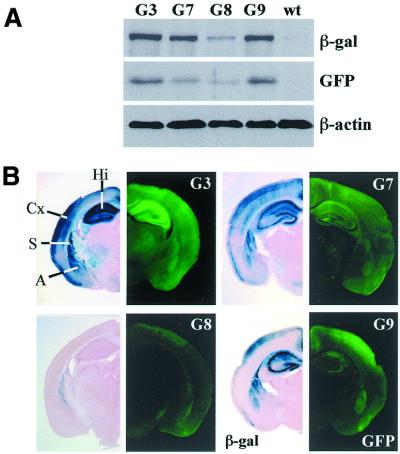
Figure 2.
GFP and β-galactosidase expression in mice derived from lines G3, 7, 8 and 9. Immunoblot analysis of GFP, β-galactosidase and β-actin in whole brain extracts of mice derived from G3, 7, 8 and 9 compared to wild-type mice (wt). Proteins were resolved on SDS–polyacrylamide gels, transferred onto a nitrocellulose membrane and probed with antibodies that recognize GFP, β-galactosidase and β-actin. Antibodies were visualized by chemoluminence method. (B) Stereomicroscopic visualization of β-galactosidase (left) and GFP (right) expression in alternate coronal brain sections of the mice derived from G3, 7, 8 and 9 at P15. The β-galactosidase staining is co-localized with GFP fluorescence in all four lines. It is restricted to the cortex (Cx, layer 2,3 and 5), hippocampus (Hi), striatum (S) and amygdala (A). Line G8 shows moderate expression and GFP fluorescence in the overview is comparable to autofluorescence of brain sections of a wild-type mouse.

