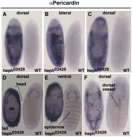
Figure 4. Pericardial cells are in excess and mislocalized in heph03429 embryos indicating that cardiogenesis is disrupted in these embryos.
A–C. The same heph03429 and wild type embryos imaged from different perspectives. D. Pericardin was present in unusual places in heph03429 embryos, such as the head and the tail. Note the large amnioserosal region (as) that persisted in heph03429 embryos. E. The ventral epidermis was reasonably developed in heph03429 embryos indicating that these embryos produced sufficient amounts of canonical Notch signaling in the ventral region. F. Pericardial cells continued to be in excess and cardiogenesis blocked in heph03429 embryos at the time when embryogenesis and dorsal vessel formation was complete in wild type embryos (stage 17). All embryos shown were from the same experiment and were processed identically.

