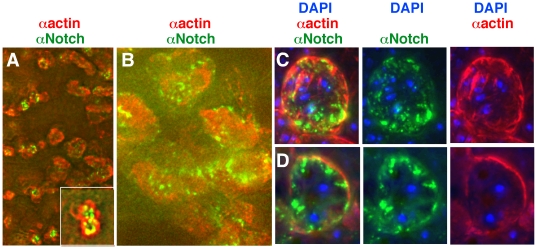
Figure 8. Notch and actin accumulated near the cell surfaces in the dorso-lateral regions of heph03429 embryos.
A, B. Low magnification images from DeltaVision restoration microscopy showing that Notch and actin expression overlap. C, D. Two sets of high magnification images from DeltaVision microscopy showing that Notch and actin accumulate near the surfaces of fused cells and not in the nucleus (marked by DAPI).

