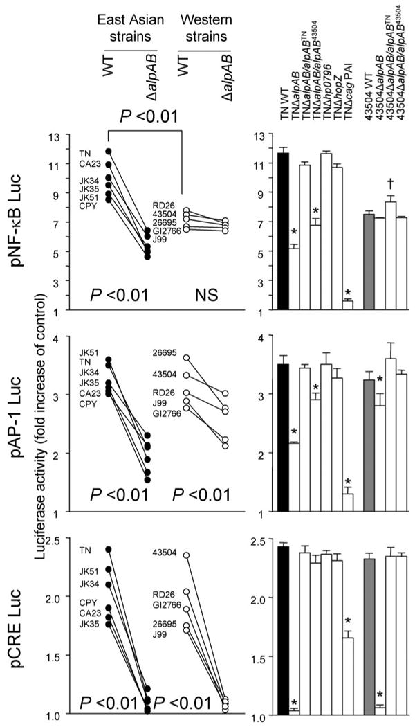
FIGURE 5. Transcriptional activity of NF-κB, AP-1, and CRE following H. pylori infection.

Three independent transfections, each done in triplicate, were performed. MKN28 cells were transiently transfected with 1.2 μg of pNF-κBluc, pAP-1luc, or pCREBluc reporter plasmids separately or together with 10 ng of Renilla plasmid as an internal control. The cells were then infected with H. pylori for 9 h. Untreated cells served as controls. Normalized luciferase activity induced by H. pylori infection is expressed as -fold increase in H. pylori-infected cells relative to uninfected controls. Data are expressed as mean (left) and mean ± S.E. (right). *, p < 0.01 decreased compared with WT strains; †, p < 0.01 increased compared with WT strains.
