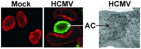
Figure 3.
The enlargement and structural changes in the nucleus and the formation of the assembly compartment during HCMV infection. The two immunofluorescence micrographs show normal nuclei in mock infected cells (left) and the significantly enlarged and kidney shaped nuclei seen in HCMV-infected cells (middle, the nuclei are visualized by staining for lamin B, red). In infected cell nuclei, a perinuclear structure known as the cytoplasmic assembly compartment or assembly complex (AC; visualized by staining for the HCMV tegument protein pp28, green) is seen nestled against the concave surface of the kidney-shaped nucleus 79–81. The immunofluorescence micrographs are reproduced from Ref. 38 with permission from the Journal of Virology. As shown in the electron micrograph (right), the AC is made of many vesicular structures; the bar indicates 2 m.

