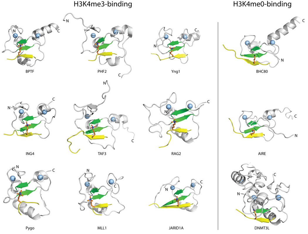
Figure 2. Structures of representative PHD fingers in complex with methylated or unmethylated H3K4 peptides.
The common elements in the binding of PHD fingers to H3 histone tails are illustrated for PHD fingers that bind H3K4me3 (i) and H3K4me0 (ii). The PHD finger structures are shown in cartoon representation with the conserved β-strands (β1 and β2) colored green, and the Zinc atoms represented by light blue spheres. The H3 peptide ligand (shown in yellow, with the K4 residue colored orange) adopts a β-strand conformation that extends the conserved anti-parallel β-sheet formed by β1 and β2 in the PHD finger. The following PDB structures were used to create this figure: 2f6j (BPTF), 3kqi (PHF2), 2jmj (Yng1), 2pnx (ING4), 2k17 (TAF3), 2v83 (RAG2), 2yyr (PYGO), 3lqj (MLL1), 2kgi (JARID1A), 2puy (BHC80), 2ke1 (AIRE), and 2pvc (DNMT3L).

