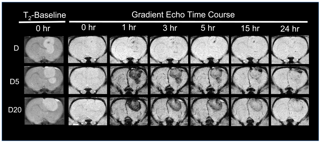
Figure 8. Visualizing post-targeting tumor clearance of D and PEG-MNPs with an MRI time course.
Immediately after magnetic targeting at 1 hour, increased hypointensity can be observed in tumors, corresponding to targeted MR images shown in Figure 5. After even a short amount of time following targeting, however, the degree of hypointensity in PEG-MNP tumors is noticeably less. Data suggest that nanoparticles begin clearance from the tumor soon after cessation of the magnetic field. It is likely that the high degree of colloidal stability possessed by PEG-MNPs minimizes nanoparticle aggregation and, thus, retention in the tumor. Agglomeration-based retention likely explains the similar tumor contrast observed at 1 and 3 hours in D targeted tumors. Coupled with histology micrographs in Figure 7, data suggest the need for additional mechanisms (e.g. tumor-specific targeting ligands) to improve tumor retention of PEG-MNPs after magnetic targeting.

