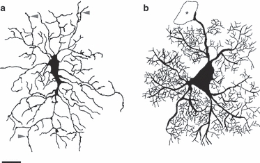
Fig. 2.

Morphological comparison between an NG2 cell and a grey matter astrocyte. Drawings of an NG2 cell (a) and an astrocyte (b) represent typical morphological appearance of the two cell types in brain slices. The drawing of the NG2 cell is traced from a Lucifer-Yellow-filled cell. Arrowheads point to varicosities present along the processes of the NG2 cell. Drawing of the grey matter astrocyte is a collage based on anti-GFP staining of astrocytes in brain slices from GFP/GFAP transgenic mouse. The asterisk indicates a blood vessel. Note the difference in the size of the soma, the thickness of the main and finer processes, as well as in the branching of fine processes. Scale bar: 10 μm (applies to both drawings). Part (a) is modified with permission from Kukley et al. (2010).
