
Fig. 2.
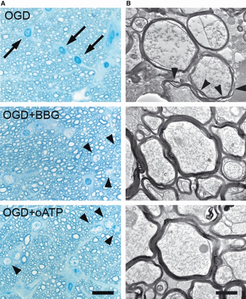
P2X7 inhibition protects myelin against ischaemic damage. (A) High-resolution analysis after 1 h oxygen-glucose deprivation (OGD) showed the presence of numerous pyknotic oligodendrocytes (arrows in left) and severe damage to myelin, with separation of the compact lamellae (arrowheads in right). In the presence of Brilliant Blue G (BBG; 50 nm) and oATP (1 mm), the number of pyknotic oligodendrocytes and the extent of myelin damage alter OGD was greatly diminished. Scale bar: 20 and 1 μm in left and right, respectively. (B) Graph summarizing the effect of BBG and oATP in oligodendroglial pyknosis and myelin damage. *P < 0.05; ***P < 0.001. Modified from Domercq et al. (2010).